Plant cells have parts that animal cells lack
Years 7–8 and 11–12Stage 4 and Stage 6Look closely
All living things are made of cells. Plant cells have a wall and a large central vacuole that animal cells lack.
Demonstration: a simplified model1 · A green plant cell
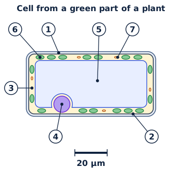
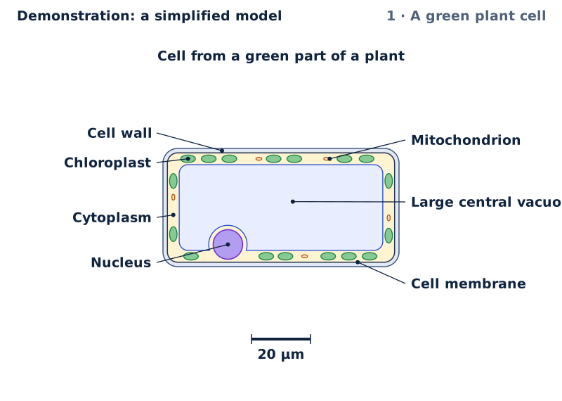
A cell from a green part of a plant: a cellulose cell wall outside the cell membrane, a large central vacuole, and chloroplasts.
- Cell wall
- Cell membrane
- Cytoplasm
- Nucleus
- Large central vacuole
- Chloroplast
- Mitochondrion
Key Cell wall: a firm layer, mostly cellulose, that supports the cell and keeps its shape. Cell membrane: controls what passes into and out of the cell. Cytoplasm: a jelly-like fluid that holds the organelles; many of the cell's reactions happen in it. Nucleus: holds the cell's DNA, the instructions that control the cell. Large central vacuole: stores water and dissolved substances; full of water, it presses outwards on the wall. Chloroplasts: use light energy to make glucose from carbon dioxide and water (photosynthesis). Mitochondria: release energy from glucose (cellular respiration). Arrows: water in the large central vacuole pressing outwards on the cell wall (turgor). Scale bar: it stands for the length written under or beside it, at the same scale as the drawing. Circle: the field of view, all that the eyepiece shows. The picture's size depends on your screen, so use the scale bar, not the magnification, to find real sizes. Iodine stains onion cells golden brown and their nuclei darker.
The model could not start here. The figure, the steps and the table on this page still show the idea; reload the page to try again.
The idea, step by step
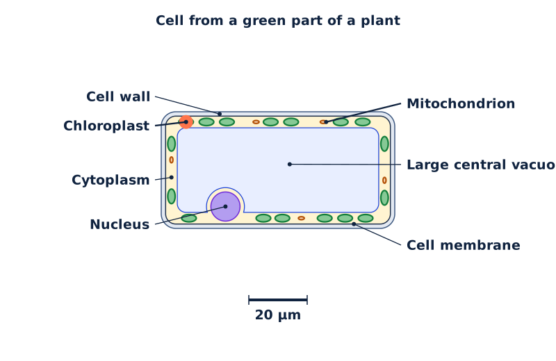
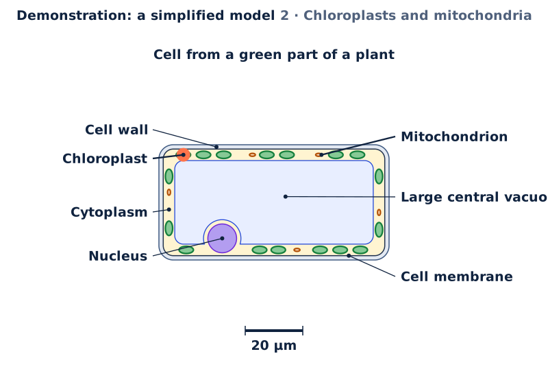
2 · Chloroplasts and mitochondriaChloroplasts use light to make glucose. Mitochondria release energy from glucose in respiration: plant cells have mitochondria too.1 Cell wall · 2 Cell membrane · 3 Cytoplasm · 4 Nucleus · 5 Large central vacuole · 6 Chloroplast · 7 Mitochondrion 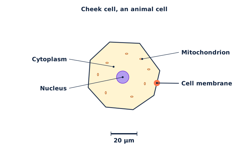
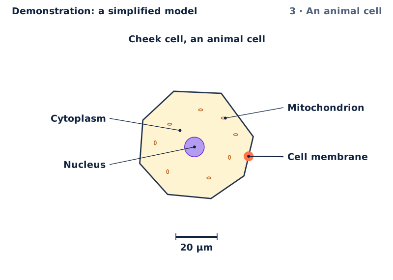
3 · An animal cellA cheek cell, an animal cell: a membrane, cytoplasm, a nucleus and mitochondria, but no cell wall, no large central vacuole, no chloroplasts.1 Cell membrane · 2 Cytoplasm · 3 Nucleus · 4 Mitochondrion 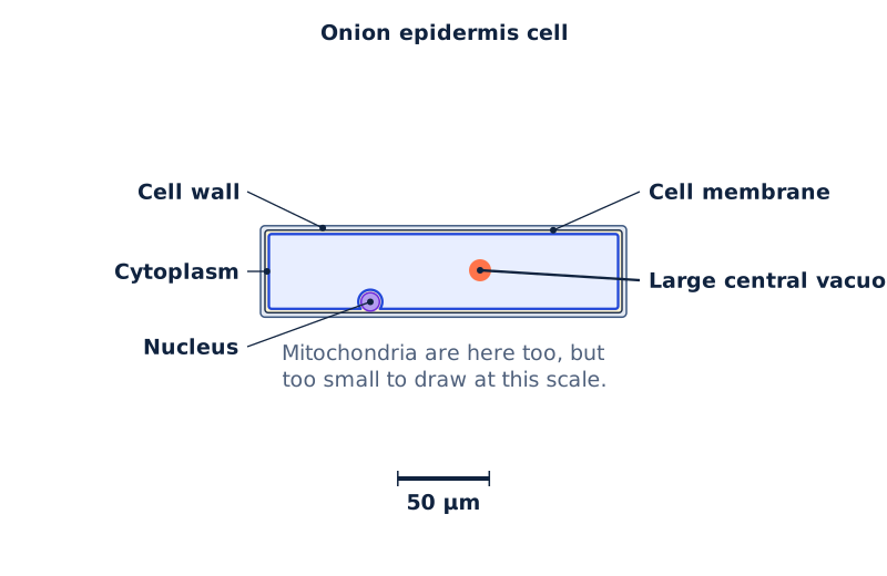
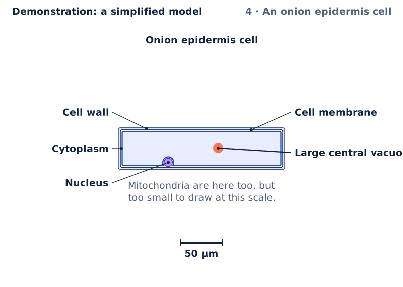
4 · An onion epidermis cellAn onion bulb's epidermis cell has a wall and a large central vacuole, but no chloroplasts: bulb scales store food and do not photosynthesise.1 Cell wall · 2 Cell membrane · 3 Cytoplasm · 4 Nucleus · 5 Large central vacuole 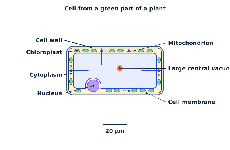
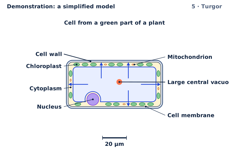
5 · TurgorFull of water, the large central vacuole presses outwards on the cell wall. This turgor keeps soft stems upright.1 Cell wall · 2 Cell membrane · 3 Cytoplasm · 4 Nucleus · 5 Large central vacuole · 6 Chloroplast · 7 Mitochondrion 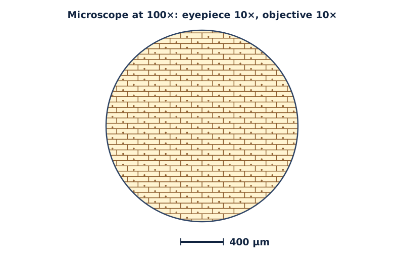
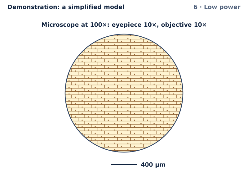
6 · Low powerLow power: a 10× eyepiece and a 10× objective give 100× in total. The field is 1800 µm across and holds many onion cells. 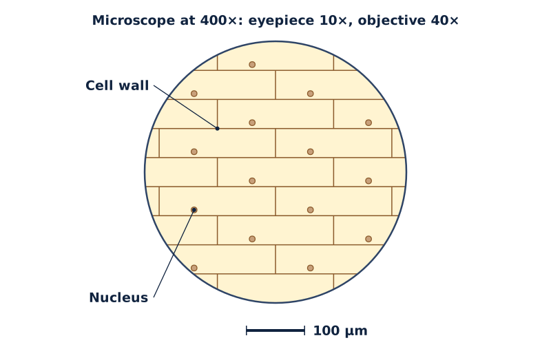
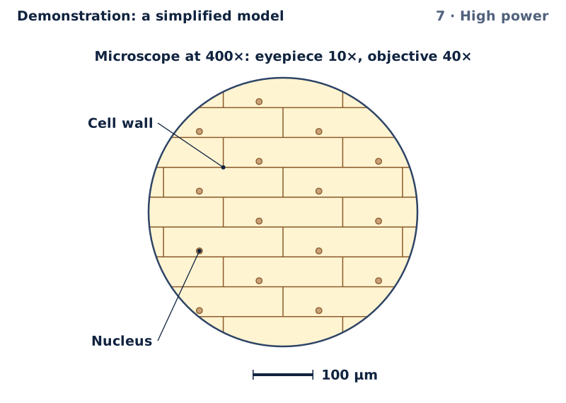
7 · High powerHigh power: the field shrinks to 450 µm, so the same bar stands for 100 µm. Each cell is still 200 µm long.1 Cell wall · 2 Nucleus


Which parts each cell has, and how long it is
| Cell | Parts it has | Parts it lacks | Real length (µm) | In the first figure |
|---|---|---|---|---|
| Green plant cell | Cell wall, cell membrane, cytoplasm, nucleus, large central vacuole, chloroplasts, mitochondria | None of these parts | 80 | Shown |
| Onion epidermis cell | Cell wall, cell membrane, cytoplasm, nucleus, large central vacuole, mitochondria | Chloroplasts | 200 | |
| Cheek cell | Cell membrane, cytoplasm, nucleus, mitochondria | Cell wall, large central vacuole, chloroplasts | 55 |
Try it in the Lab
Practicals with real materials, each with its safety card.
- Plant and animal cells: onion epidermis and cheek cells side by sideYear 8Bench practicalMedium riskSchool laboratory, not for home
- Measuring with a microscope: field of view, magnification and a scale barYear 8Bench practicalLow risk
- Prokaryotic and eukaryotic cells under the light microscope, with calibrated scale barsYear 11Bench practicalLow risk
- Measuring cells with a calibrated eyepiece graticuleYear 11Bench practicalMedium riskSchool laboratory, not for home
- Unicellular, colonial and multicellular organisms in pond water by the hanging-drop methodYear 11Bench practicalLow risk
With a learner
Three questions to ask
- Which parts do all three cells share?
- Why do the cells of an onion bulb's skin have no chloroplasts?
- At high power, has the cell grown, or has the field of view shrunk?
What to expect
Many learners say every plant cell has chloroplasts, or that plant cells have no mitochondria. Many also think a higher power shows more of the slide.
What to try next
Try a practical in Try it in the Lab, above: look at onion and cheek cells, then measure the field of view and work out a cell's real size.
About this model
What is simplified
- The cells' outlines, nuclei, chloroplasts and mitochondria are drawn to scale from the sizes listed here. Cell walls, membranes and the onion cell's thin layer of cytoplasm are drawn thicker than to scale, so that you can see them.
- Each diagram has its own scale, given by its scale bar: the onion cell is drawn at a smaller scale than the other two, because it is several times longer.
- The onion cell's mitochondria would be dots too small to draw at its scale, so they are not drawn. The table and the words say it has them.
- The diagrams show one slice through the middle of each cell. Chloroplasts also line the upper and lower surfaces of a green cell, and an animal cell can hold small vacuoles; the model does not draw them.
- Under the microscope, the model shows only the outlines, nuclei and chloroplasts that a school light microscope shows at each power. Membranes and mitochondria are too thin or too fine for it to show clearly without special stains.
- At low power the green cells' chloroplasts are too small to draw one by one, so a green fill stands for them.
- The field of view is worked out from the eyepiece's field number, listed with the numbers here. Your microscope's field number may differ, and so will its field.
- Real cells vary from one to the next: onion epidermis cells change from one layer of the bulb to another, and no two cheek cells look alike. The model draws one typical cell of each kind.
Numbers and their sources
- 10× The eyepiece of a school microscope. Total magnification is the eyepiece's magnification times the objective's. Source: Nikon MicroscopyU, Field of View (a 10x eyepiece and its field number); total magnification as eyepiece times objective, as the linked Lab practical on field of view and scale sets it, read 26 September 2026.
- 10× Low power in the model is this objective. Source: Nikon MicroscopyU, Useful Magnification Range, Table 1 (the common 10x and 40x objectives), read 26 September 2026.
- 40× High power in the model is this objective. Source: Nikon MicroscopyU, Useful Magnification Range, Table 1 (the common 10x and 40x objectives), read 26 September 2026.
- 18 mm The eyepiece's field number. The field of view on the slide is the field number divided by the objective's magnification, so it shrinks as the power rises. Microscopes differ: check the number on your eyepiece. Source: Nikon MicroscopyU, Field of View: field of view = field number / objective magnification; a 10x eyepiece typically has a field number of 16 to 18 mm, read 26 September 2026.
- 200 µm Onion epidermis cells differ in size from one layer of the bulb to the next. The model draws one of a typical length. Source: Science and Plants for Schools (SAPS), Calibrating and measuring in microscopy: exploring onion growth (cell size changes between the layers of the bulb), read 26 September 2026.
- 50 µm A typical width of an onion epidermis cell, measured the same way. Source: Biology 100 laboratory key, Cells: onion epidermal cells about 40 to 50 micrometres wide from the field of view, read 26 September 2026.
- 80 µm The green cell is drawn from a photosynthetic cell of this size, whose volume is mostly a large vacuole, with its cytoplasm and chloroplasts at the edge. Source: Milo and Phillips, Cell Biology by the Numbers, How large are chloroplasts? (lamina cells about 80 micrometres long and 40 wide), read 26 September 2026.
- 40 µm The green cell's width. Source: Milo and Phillips, Cell Biology by the Numbers, How large are chloroplasts?, read 26 September 2026.
- 55 µm Cheek cells are flat and irregular. Measured healthy cheek cells averaged just over this; the model rounds up. Source: Cytological and Cytomorphometric Study of Exfoliated Cells of the Oral Mucosa in Diabetic Patients (2020), Table 2, control group: cell diameter 53.1 micrometres, nuclear diameter 9.5 micrometres, read 26 September 2026.
- 10 µm Nuclei are typically a few micrometres across, up to this size; the plant cells' nuclei are drawn at the upper end. Source: Milo and Phillips, Cell Biology by the Numbers, How big are nuclei? (typical diameters between 2 and 10 micrometres), read 26 September 2026.
- 9.5 µm The cheek cell's nucleus. Source: Cytological and Cytomorphometric Study of Exfoliated Cells of the Oral Mucosa in Diabetic Patients (2020), Table 2, control group, read 26 September 2026.
- 5 µm Chloroplasts in plants are lens-shaped, about this size across. Source: Milo and Phillips, Cell Biology by the Numbers, How large are chloroplasts? (a characteristic diameter of about 4 to 6 micrometres), read 26 September 2026.
- 2 µm A mitochondrion is drawn as its textbook shape, about this long. Source: Milo and Phillips, Cell Biology by the Numbers, How big are mitochondria? (a length of roughly two microns and a diameter of roughly one), read 26 September 2026.
- 1 µm A mitochondrion's width. Source: Milo and Phillips, Cell Biology by the Numbers, How big are mitochondria?, read 26 September 2026.
Review
Demonstration: a simplified model. Checked against its written sources, 26 September 2026. Not reviewed by a qualified teacher.
Curriculum references
SC4-CLS-01SC4-WS-01BI-11-01BI-11WS-03BI-11WS-04BIO11-8BIO11/12-3BIO11/12-4AC9S8U01AC9S8I03AC9S8I04
Reference, not a verified alignment.